Multi-frequency Harmonic Acoustography for Tissue Identification and Border Detection

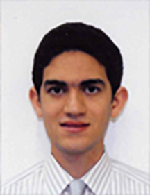
Speaker: Ashkan Maccabi
Affiliation: Ph.D. Candidate - UCLA
Abstract: Tissue and border detection of diseased regions are essential for surgeons during surgical operations in tumor excisions. In the absence of an imaging technique that provides quantitative well-defined margins by separating diseased from healthy regions, palpation of the tissue during surgery is the only practical option. To ensure the complete resection of the diseased region, the surgeon is inclined to have large margins surrounding the site. The goal of this project is to develop an imaging modality that utilizes vibroacoustography (VA) technique to spatially map the contrast in mechanical and acoustic properties between malignant and normal tissue for abnormal detection. By enhancing boundary regions of malignant and healthy tissue by this novel imaging technique, margin around sites are decreased and as a result, the resection of healthy tissue is minimized. Some approaches that are used in the detection of tumor regions include conventional ultrasound, manual palpation, and CT scan. However, they suffer from limitations such as lack of sensitivity, subjectivity, and low contrast. As an alternative, VA provides an enhanced image of the boundary using the mechanical and acoustic properties of the targets as the main contrast mechanism. This work outlines the development of a VA medical imaging system for enhanced border detection:
- Construction, development, and advancement of the VA system
- Characterization and optimization of VA system parameters such as point spread function (PSF), lateral and axial resolution of the imaging beam, sensitivity and specificity specifications for biomedical imaging applications
- Investigation of VA feasibility in imaging mechanical properties of targets
- Design of a compact VA system for in vivo applications to enhance tissue characterization and boundary detection
The long-term goal of VA medical imaging is to provide a quantitative method to detect the border between malignant and healthy tissue intra-operatively during surgical procedures.
Biography: Ashkan Maccabi received his B.S. degree in Bioengineering and M.S. degree in Biomedical Engineering from University of California, Los Angeles (UCLA) in 2011 and 2013, respectively. He joined Professor Grundfest’s Biophotonics laboratory in 2010 as an undergraduate researcher focusing on medical imaging. Mr. Maccabi began his work in acoustics in the fall of 2011 at UCLA. He has published over 10 journal articles and conference proceedings in the field of THz and novel ultrasound system developments. He has attended multiple conferences including BMES, SPIE BiOS, MMVR, SPIE Medical Imaging, UC System wide Symposium, and IEEE EMBC to present his research on vibroacoustography. His research interests include the development of a novel dual-frequency ultrasound technique for measurements of tissue viscoelastic properties for tissue identification and border detection. He has been a mentor to more than 25 undergraduates and high school students interested in STEM programs under HSSRP and CEED, throughout all five years of his graduate study.
For more information, contact Prof. Warren Grundfest (warrenbe@seas.ucla.edu)
Date/Time:
Date(s) - Oct 19, 2016
4:30 pm - 6:30 pm
Location:
E-IV Tesla Room #53-125
420 Westwood Plaza - 5th Flr., Los Angeles CA 90095
