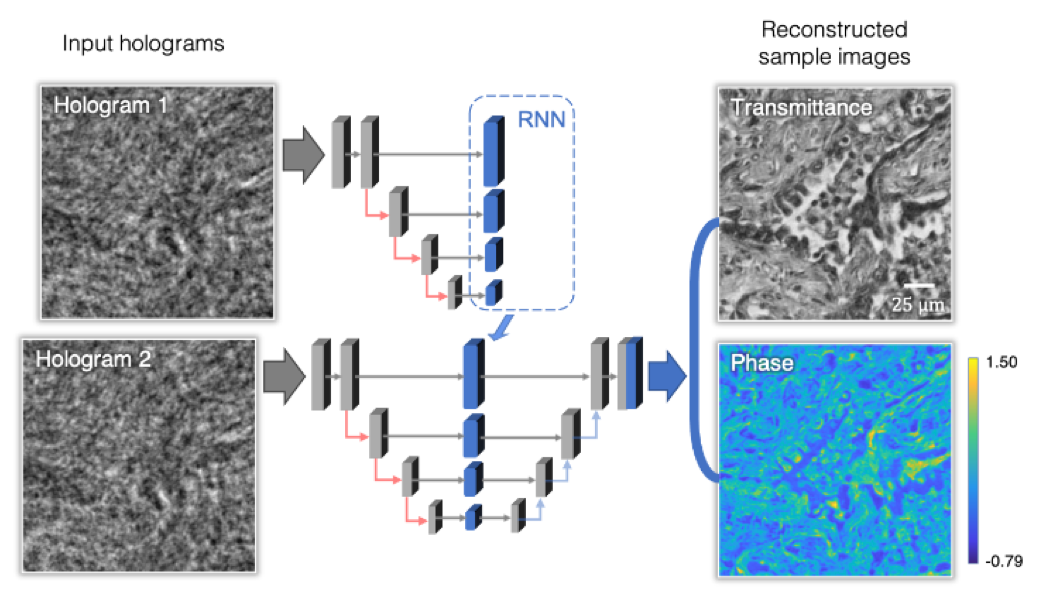
Figure Caption: UCLA researchers demonstrated holographic imaging using recurrent neural networks (RNN). A human lung tissue section is imaged with faster and better hologram reconstruction.
Digital holographic imaging is a commonly used microscopy technique in biomedical imaging that reveals rich optical information of the sample, which could be used, for example, to detect pathological abnormalities in tissue slides. Common image sensors only respond to the intensity of the incoming light. Therefore, reconstructing the complete 3D information of a hologram that is digitally recorded by such sensors has been a challenging task involving optical phase retrieval, which is a time-consuming and computationally intensive step in digital holography.
A research team at UCLA has recently developed a novel holographic phase retrieval technique that can rapidly reconstruct microscopic images of samples with up to 50-fold acceleration compared to existing methods. This new technique is taking advantage of recurrent neural networks trained using deep learning and incorporates spatial features from multiple holograms to digitally create holographic microscopy images of samples, such as human tissue slides. This results in better image quality and faster reconstruction speed, while also enhancing the depth-of-field of the reconstructed sample volume.
This work was published in ACS Photonics, a journal of the American Chemical Society. UCLA researchers showed the effectiveness of this new holographic imaging method through experiments performed on human lung tissue sections and Pap smears, commonly used for screening of cervical cancer. These results report the first demonstration of the use of recurrent neural networks for holographic imaging and phase recovery, and also open up new opportunities for designing improved holographic microscopes with reduced number of measurements and increased imaging speeds.
“This framework can be broadly applicable to various biomedical imaging modalities, including for example fluorescence microscopy, to efficiently utilize a sequence of acquired images to rapidly and accurately create 3D reconstructions of a sample volume,” said Dr. Aydogan Ozcan, the Chancellor’s Professor of Electrical and Computer Engineering at UCLA and an associate director of the California NanoSystems Institute, who is the senior corresponding author of the work.
The other authors include graduate students Luzhe Huang, Tairan Liu, Xilin Yang, Yi Luo and Professor Yair Rivenson, all from the Electrical and Computer Engineering department at UCLA. Professor Ozcan also has UCLA faculty appointments in bioengineering and surgery, and is an HHMI professor.
Link to paper: https://pubs.acs.org/doi/abs/10.1021/acsphotonics.1c00337