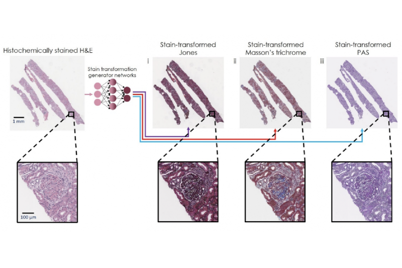
Figure Caption: Virtual transformation and re-staining of one tissue biopsy stain (H&E) into three special stains using deep neural networks. Image credit: Ozcan Lab @ UCLA.
In order to perform medical diagnoses, pathologists visually inspect histochemically stained tissue biopsy sections. The hematoxylin and eosin (H&E) stain is the most used histochemical stain in pathology, covering the majority of the human tissue biopsy stains performed globally. However, in many clinical cases, additional “special stains” are needed to provide contrast and color to different tissue components and allow pathologists to get a clearer diagnostic picture. These special stains often require significantly longer tissue preparation time, along with laborious effort and monitoring by expert histotechnologists, all of which increase the costs and time to diagnosis.
Researchers at UCLA developed a deep learning-based technique which can be used to eliminate the need for these special stains to be prepared by human histotechnologists, by computationally transforming existing images of the H&E stained tissue into special stains. This AI-based technique was demonstrated by generating a full panel of special stains used for kidney tissue, namely, Periodic acid–Schiff (PAS), Jones silver stain, and Masson’s Trichrome; all of these special stains were computationally transformed, using specialized deep neural networks, from existing images of H&E stained tissue biopsies. The researchers performed a clinical evaluation using this panel of special stains to demonstrate the efficacy of this stain-to-stain transformation technique on a variety of clinical samples, covering a broad range of kidney diseases. This evaluation performed by a multi-institution team of board-certified renal pathologists found a statistically significant improvement in the diagnoses that were achieved by using the neural network generated special stains and the H&E images over the use of the H&E images only. An additional study also showed that the quality of the virtually re-stained images is statistically equivalent to those which were histochemically stained by human experts.
This stain-to-stain transformation is fast, taking less than 1 minute for a needle core tissue biopsy section. This speed enables it to improve the quality of preliminary diagnoses, where special stains are needed, also providing significant time and cost savings. These advantages are particularly important when diagnosing medical conditions such as transplant rejection cases, where a fast and accurate diagnosis enables rapid treatment which may lead to significantly improved clinical outcomes. Furthermore, since the virtual re-staining technique is applied to existing stains, it is easy to adopt as it does not require any changes to the current tissue processing workflow used in pathology.
This research, published in the journal Nature Communications, was led by Dr. Aydogan Ozcan, the Chancellor’s Professor of Electrical and Computer Engineering at UCLA, and an associate director of the California NanoSystems Institute (CNSI), Dr. W. Dean Wallace, a Professor of Pathology at the USC Keck School of Medicine, and Dr. Yair Rivenson, an adjunct Professor of Electrical and Computer Engineering at UCLA, along with UCLA graduate students, Kevin de Haan, Yijie Zhang, and Tairan Liu. Clinical validation of this virtual re-staining method was advised by Dr. Jonathan Zuckerman from the UCLA Department of Pathology and Laboratory Medicine at the David Geffen School of Medicine.
The research was supported by the NSF Biophotonics Program.
Link to the published paper: https://rdcu.be/ctUXo
Link to Ozcan Lab at UCLA: https://innovate.ee.ucla.edu/